RCMIA (Rare Cell Multiprobe Imaging Analyser) revolutionizes the microscopic imaging of rare cells. With high-resolution scans and the analysis of up to 50 epitopes, this technology is ideal for the cytology of hypocellular fluids or for immunophenotyping.
Suitable Applications: • Cytological analysis of human or animal hypocellular fluids (CSF, urine, peritoneal fluid, etc.) or other samples containing valuable rare cellular materials • Comprehensive immunophenotyping • High-throughput analysis / iterative investigation of ex-vivo samples (optional storage of samples after initial analysis for extended studies over several weeks)
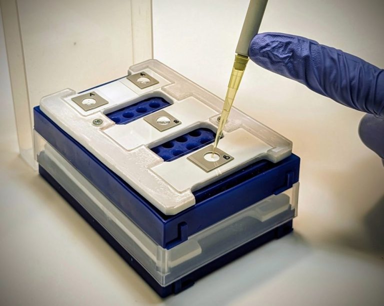
Preparation of Thin Cell Layers
• Cavities with 2, 4, 8, 12 mm diameters • Antibody-assisted cell enrichment • Covalent cell fixation if required
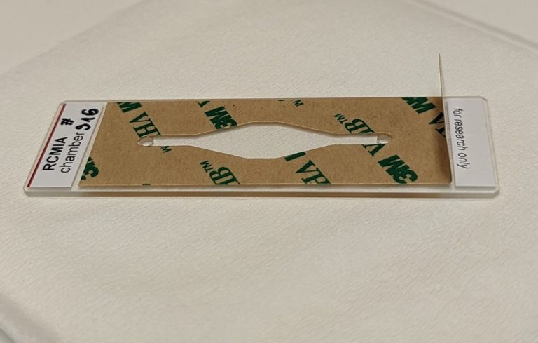
Staining Chambers
• Convenient and secure sample storage • Long-term preservation over several months • Ready-to-use for microfluidic staining
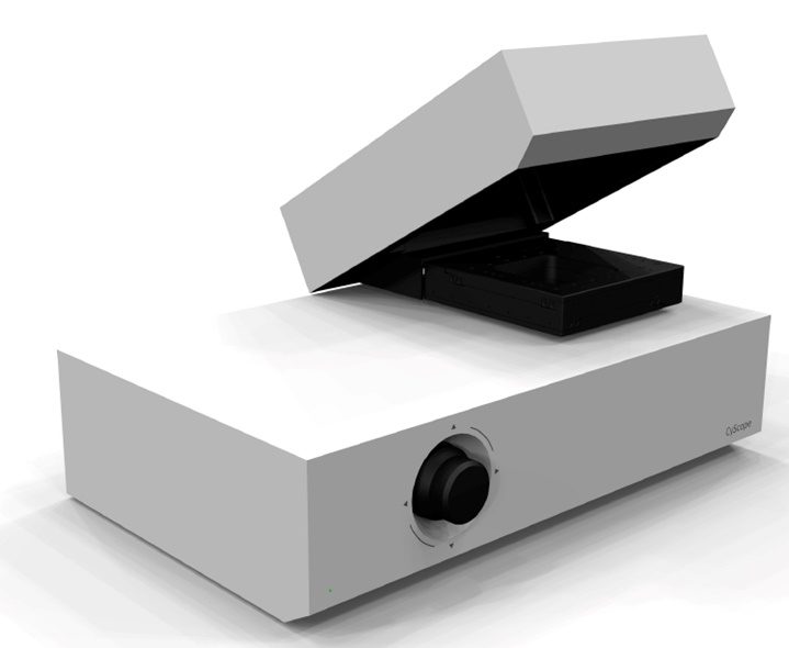
Microscopic Imaging
• Large scanning areas (100 mm² and more) • 4 fluorescence channels (UV, FITC, PE, APC) + brightfield • Excellent resolution • Comprehensive image analysis
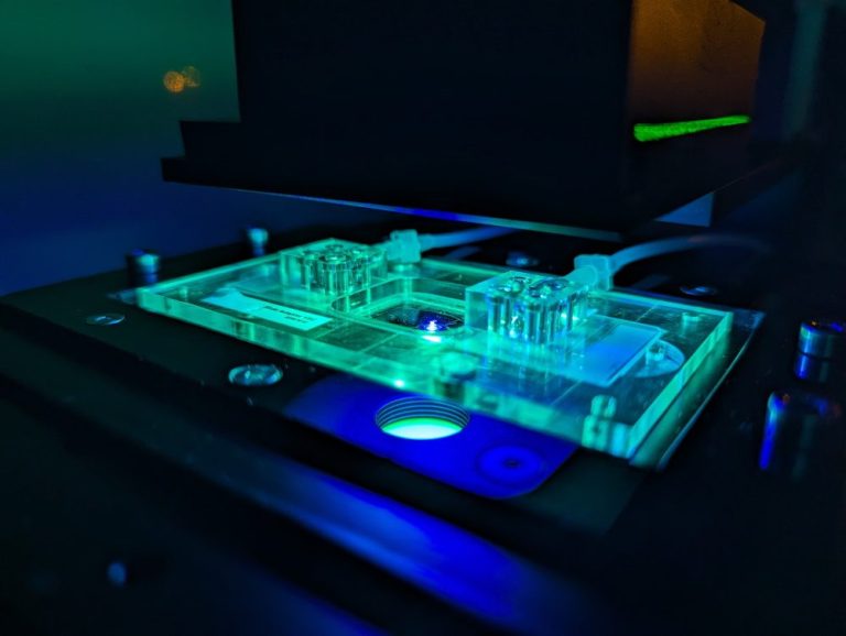
Sequential Immunostaining
• Up to 20 staining and bleaching cycles
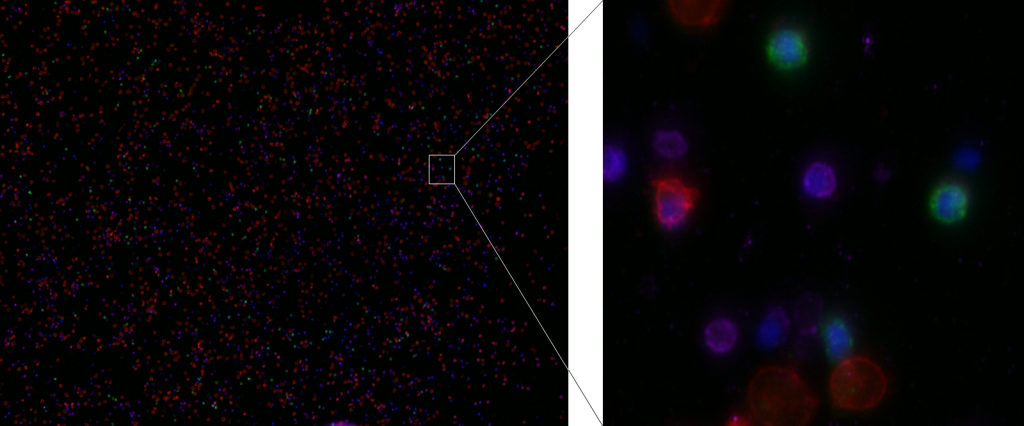
Cell staining of human leukocytes with DAPI, CD14-Atto488, CD16-PE, CD3-APC
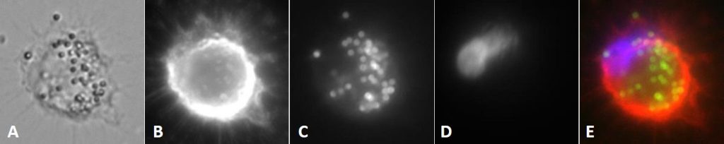
Microscopic image of a THP1 cell with 1 µm beads. A: Brightfield image, B: CD45-PE, C: Atto488 beads, D: DAPI, E: Overlay of fluorescence channels
Funding and Collaboration
Developed within the NADIM project funded by the German Federal Ministry of Education and Research (BMBF), this technology has been advanced in close collaboration with Invigate GmbH, Quantum Analysis GmbH, and Cytecs GmbH.